
Eye Coloboma: A Clinician-Level Guide to Embryology, OCT Findings, Retinal Detachment Risk, and Management

What Is an Eye Coloboma?
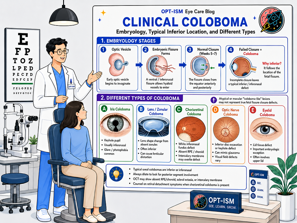
An ocular coloboma is a congenital defect caused by incomplete closure of the embryonic fissure, also called the fetal fissure, choroidal fissure, or optic fissure. It may involve the iris, ciliary body, zonules, lens contour, choroid, retina, optic nerve, or a combination of these structures. Typical uveal colobomas are classically located in the inferior or inferonasal portion of the involved structure.
Clinically, coloboma can range from a subtle inferonasal iris sphincter notch to a large chorioretinal defect involving the optic nerve and macula. Visual prognosis depends less on the cosmetic appearance of the iris and more on whether the macula, optic nerve, foveal architecture, refractive status, amblyopia, retinal detachment, or choroidal neovascularization are involved. A comprehensive clinical review notes that vision is affected when the disc or fovea is involved or when complications such as retinal detachment, CNV, cataract, or amblyopia occur.
Embryology: Why Colobomas Happen
During early ocular development, the optic vesicle invaginates to form the double-layered optic cup. A ventral groove forms along the optic cup and optic stalk. This groove is the embryonic fissure. Its purpose is not accidental: it allows mesenchymal tissue and the hyaloid vasculature to enter the developing eye. The fissure normally closes around the fifth to seventh week of gestation.
Closure begins near the equator and proceeds both anteriorly and posteriorly. Failure of anterior closure can produce iris, ciliary body, zonular, or lens-related findings. Failure of posterior closure can produce chorioretinal or optic nerve coloboma.
This embryologic timeline explains why a patient may have an isolated iris coloboma, an isolated posterior segment coloboma, or a “complete” coloboma extending from the iris to the optic nerve. A complete coloboma may involve the iris, ciliary body, zonules, lens contour, choroid, retina, and optic disc.
Why Are Colobomas Inferior?
The simplified teaching is that “colobomas are inferior,” but the more precise statement is:
Typical uveal colobomas are inferior or inferonasal because the embryonic fissure is located on the ventral/inferonasal aspect of the developing optic cup.
The embryonic fissure forms from eccentric invagination of the optic vesicle, leaving an inferonasal gap that later closes. When closure fails, the remaining defect follows that embryologic line, so typical iris, chorioretinal, and optic nerve colobomas are found inferiorly or inferonasally.
In optic nerve coloboma, the fissure closes ventrally last, so the defect is usually ventral/inferior.
However, clinicians should avoid saying that all colobomas are inferior. Typical embryonic fissure colobomas are inferior/inferonasal. But “atypical colobomas” may be described in other locations, and some lesions historically called “macular colobomas” may not be true embryonic fissure closure defects. Macular coloboma-like lesions can be developmental, genetic, inflammatory, or infectious scars, including congenital toxoplasmosis.
There is also an important eyelid exception. Eyelid colobomas do not share the same embryologic origin as uveal colobomas and are often located at the superior medial upper eyelid.
Clinical Types of Coloboma
Iris Coloboma
Iris coloboma classically presents as an inferonasal keyhole-shaped pupil. Patients may report glare, photophobia, monocular diplopia, or visual distortion. Cosmetic concerns are also common. Iris coloboma may coexist with ciliary body, zonular, chorioretinal, or optic nerve coloboma, so the posterior segment must be examined carefully.
Clinical pearls:
Always dilate if an iris coloboma is seen.
Look for inferior lens notching or zonular absence.
Ask about photophobia and glare, not just visual acuity.
Consider prosthetic/tinted contact lenses for glare control.
Differentiate congenital iris coloboma from traumatic iris loss or surgical iridectomy.
Lens and Zonular “Coloboma”
A “lens coloboma” is technically a misnomer. The lens tissue itself is not usually missing. Instead, absent or abnormal zonules create localized flattening or notching of the lens equator. This is often seen inferiorly in the same embryologic axis as the iris or ciliary body defect.
Clinical pearls:
Look carefully after dilation.
Expect irregular astigmatism or lenticular distortion.
Be cautious with cataract surgery planning because zonular support may be abnormal.
In extensive coloboma or microphthalmia, biometry can be challenging.
Chorioretinal Coloboma
Chorioretinal coloboma appears as a sharply demarcated white or pale defect, often with scleral show, scalloped borders, and pigment at the margins. The choroid and RPE are absent within the colobomatous area, and the overlying thin retinal tissue is often referred to as the intercalary membrane.
The fundus appearance varies widely. Small incomplete lesions may appear as inferonasal RPE/chorioretinal atrophy, posterior dipping of the ora, or subtle changes along the line between the disc and inferonasal periphery. Larger defects may extend from the inferior periphery toward or through the optic disc and macula.
Optic Nerve Coloboma
Optic nerve coloboma is typically a sharply demarcated inferior excavation of the optic nerve head. It may mimic glaucomatous cupping, especially if the clinician focuses only on cup size rather than congenital disc architecture. A typical optic disc coloboma has a bowl-shaped excavation, often occupying the lower part of the disc, without the central glial tuft and radial vessel pattern seen in morning glory disc anomaly.
Clinical pearls:
Do not diagnose glaucoma based on cup size alone.
Compare optic nerve appearance with IOP, corneal thickness, gonioscopy, OCT, and visual field.
Expect visual field defects that may not respect typical glaucomatous patterns.
Baseline fundus photography is extremely helpful.
Consider systemic evaluation when bilateral or associated with developmental abnormalities.
OCT Findings in Coloboma
OCT is useful for documenting anatomy, identifying complications, and distinguishing coloboma from look-alike conditions.
Chorioretinal Coloboma OCT
On OCT, chorioretinal coloboma may show absence or severe thinning of RPE and choroid, scleral excavation or ectasia, and an overlying intercalary membrane. OCT can help identify subtle breaks in the intercalary membrane, subclinical retinal detachment, retinoschisis-like separation, or subretinal/intraretinal fluid.
Incomplete fundus colobomas may show mild ectasia with relatively preserved retinal architecture, whereas smaller true colobomatous defects may show a hyperreflective membrane without normal retinal layering.
Optic Nerve Coloboma OCT
OCT of optic nerve coloboma may demonstrate retinochoroidal-scleral excavation of the nerve. OCT can also help separate optic nerve coloboma from optic pit, morning glory disc anomaly, peripapillary staphyloma, and glaucomatous cupping.
Interpretation warning: RNFL and GCC printouts can be misleading in congenital anomalous discs. Segmentation errors, abnormal disc size, excavation, and atypical anatomy may produce abnormal-looking maps that do not necessarily represent progressive glaucoma.
OCT-A and CNV
OCT and OCT-A may help detect choroidal neovascularization, especially near the coloboma margin. CNV in chorioretinal coloboma is often located at the border of the defect, where Bruch’s membrane/RPE disruption and pigmentary change may exist.
Retinal Detachment Risk
The most important posterior segment complication of chorioretinal coloboma is retinal detachment. Reported retinal detachment rates vary widely across hospital-based studies, with one clinician review reporting a range from 2.4% to 47.5%.
A pediatric chorioretinal coloboma study reported retinal detachment in 17.6% of eyes. The same study found a lower detachment prevalence in eyes treated with prophylactic laser photocoagulation compared with untreated eyes, 2.9% versus 24.1%, suggesting a protective effect in selected cases.
Another series of chorioretinal coloboma complications found retinal detachment in 29.4% of eyes and CNV in 13.7%. Importantly, retinal breaks were not always inside the coloboma. Some were outside the coloboma, supporting the idea that vitreoretinal interface abnormalities and peripheral retinal pathology also contribute to detachment risk.
Mechanisms of retinal detachment may include:
Breaks in the thin intercalary membrane
Breaks at the coloboma margin
Retinal breaks outside the coloboma
Abnormal vitreoretinal traction
Communication between abnormal retinal, subretinal, and colobomatous spaces
Associated optic nerve cavitary anomalies
This is why a careful peripheral retinal exam matters even when the coloboma itself appears stable.
Should Colobomas Receive Prophylactic Laser?
Prophylactic laser around a chorioretinal coloboma is a nuanced retina decision. It may reduce detachment risk in selected eyes, but complete treatment can be difficult when the optic disc lies within or adjacent to the coloboma. A major review notes that prophylactic laser to the coloboma border appears attractive for reducing detachment risk, but many eyes have the optic disc within the choroidal coloboma, making complete safe treatment difficult.
Clinical approach:
Refer to retina if there is a large posterior segment coloboma, suspicious border changes, subretinal fluid, symptoms, or poor view.
Educate patients about RD symptoms: flashes, floaters, curtain/shadow, sudden vision change.
Consider baseline widefield photos.
Do not assume “stable since birth” means “no lifetime RD risk.”
In children, exam under anesthesia may be needed if adequate peripheral evaluation cannot be obtained.
Differential Diagnosis
Iris Coloboma Differential
Traumatic iris defect
Surgical iridectomy or peripheral iridotomy
Essential iris atrophy/ICE spectrum
Aniridia or partial aniridia
Corectopia
Posterior synechiae causing irregular pupil
Iris melanoma or sectoral iris atrophy
Congenital iris coloboma typically has smooth margins and an inferonasal location. If the cornea is normal and the defect looks atypical, look carefully for prior surgery or trauma.
Chorioretinal Coloboma Differential
Congenital toxoplasmosis scar
Macular coloboma-like atrophic scar
Myopic chorioretinal atrophy
Chorioretinal scar from infection or inflammation
Morning glory disc anomaly
Peripapillary staphyloma
Optic disc pit with maculopathy
Tilted disc syndrome
Staphylomatous myopic degeneration
Retinal pigment epithelium hypertrophy or reactive pigmentation at coloboma margin
Choroidal melanoma when pigmentation or elevation is atypical
Macular coloboma deserves special caution. A lesion called “macular coloboma” may not be related to embryonic fissure closure and can represent congenital infection, retinal dystrophy, or another developmental/inflammatory scar.
Optic Nerve Coloboma Differential
Morning glory disc anomaly
Optic disc pit
Peripapillary staphyloma
Optic nerve hypoplasia
Megalopapilla
Tilted disc
Glaucomatous cupping
Congenital cavitary optic disc anomaly
Morning glory disc anomaly usually has a funnel-shaped excavation, central glial tuft, peripapillary pigmentary change, and radial retinal vessels. Optic nerve coloboma usually lacks the central glial tuft and has more normal vasculature aside from displacement by the excavation.
Systemic and Genetic Associations
Many colobomas are isolated and sporadic, but systemic associations must be considered, especially in bilateral disease, microphthalmia, developmental delay, hearing loss, craniofacial anomalies, cardiac findings, renal disease, or growth abnormalities.
Important associations include:
CHARGE syndrome
PAX2-related renal-coloboma syndrome
Cat eye syndrome
Trisomy 13 or 18
Goldenhar syndrome
Joubert syndrome
SOX2-related eye disorders
Meckel syndrome
Rubinstein-Taybi syndrome
Warburg/Walker-Warburg spectrum
CHARGE syndrome is one of the classic associations and includes coloboma, heart defects, choanal atresia, growth/developmental delay, genital abnormalities, and ear/hearing anomalies.
A Moran CORE review notes that ocular colobomas are often associated with systemic disorders and reports CHARGE syndrome as a common association in ocular coloboma cohorts.
Clinical Workup
A practical clinician-level evaluation should include:
History
Age at diagnosis
Unilateral vs bilateral
Family history of coloboma, congenital cataract, renal disease, hearing loss, developmental delay
Prematurity or prenatal exposures
Prior trauma or eye surgery
Symptoms of glare, photophobia, diplopia, reduced vision, field loss
Flashes, floaters, shadow, curtain, or sudden acuity change
Pediatric developmental history and school performance
Examination
Best-corrected visual acuity
Cycloplegic refraction in children
Pupils and APD assessment
Ocular alignment and motility
Nystagmus assessment
Slit lamp exam of iris, lens, zonules, and cornea
IOP and gonioscopy when indicated
Dilated fundus exam with careful peripheral retina evaluation
Widefield fundus photography
OCT macula and optic nerve
OCT through the coloboma margin when possible
Visual field testing when age-appropriate
B-scan if poor view or suspected staphyloma/cyst
Referral Considerations
Refer to retina for large posterior segment coloboma, subretinal fluid, suspected break, CNV, symptomatic floaters/flashes, or difficult peripheral assessment.
Refer to pediatric ophthalmology for infants/children, amblyopia risk, strabismus, nystagmus, or poor cooperation.
Consider genetics, pediatrics, ENT, cardiology, nephrology, or neurology when bilateral coloboma, microphthalmia, developmental delay, hearing concerns, craniofacial anomalies, renal concerns, or CHARGE/PAX2 features are present.
Management Pearls for Eye Doctors
The iris finding is not the whole diagnosis.
An iris coloboma should trigger a dilated exam to look for ciliary body, lens, chorioretinal, or optic nerve involvement.
Inferior/inferonasal location supports a typical embryonic fissure coloboma.
Superior, temporal, or macular lesions should prompt a broader differential.
Do not overcall glaucoma in optic nerve coloboma.
Congenital excavation can mimic cupping. Look for progression, IOP risk, rim change, disc hemorrhage, corneal thickness, angle findings, and repeatable visual field/OCT change.
OCT RNFL printouts may be unreliable.
Congenital disc anomalies often produce segmentation artifacts and abnormal normative database comparisons.
Retinal detachment counseling is essential.
Patients should know the symptoms of RD and understand that risk can persist lifelong.
Baseline imaging matters.
Widefield photos, OCT, and visual fields provide a reference point for future change.
Think amblyopia early.
In children, refractive correction, anisometropia management, patching/atropine therapy, and strabismus care may be time-sensitive.
Protect the better-seeing eye.
If one eye has poor visual potential, recommend polycarbonate lenses and sports eye protection.
Manage glare.
Iris coloboma patients may benefit from tinted lenses, polarized sunglasses, prosthetic contact lenses, or artificial pupil designs.
Do not ignore systemic clues.
Bilateral coloboma, microphthalmia, developmental delay, hearing loss, renal findings, or craniofacial anomalies should prompt systemic evaluation.
Patient Counseling Language
A helpful way to explain this to patients:
“Coloboma means part of the eye did not fully form before birth. In many people it stays stable, but the effect on vision depends on which part of the eye is involved. When it affects only the iris, the main issue may be glare or appearance. When it affects the retina or optic nerve, it can affect vision more significantly and may increase the risk for retinal complications. The goal of monitoring is to protect vision, watch for complications early, and make sure children develop the best vision possible.”
Key Takeaway
Typical ocular colobomas are inferior or inferonasal because they follow the embryologic location of the fetal fissure. The most important clinical questions are not simply “Is the pupil keyhole-shaped?” but rather:
Is the macula involved?
Is the optic nerve involved?
Is there amblyopia risk?
Is there retinal detachment or CNV risk?
Is this isolated or syndromic?
Is the finding truly a typical coloboma, or a look-alike lesion?
For clinicians, the best management strategy is careful diagnosis, baseline imaging, risk-based follow-up, early amblyopia care, retinal detachment education, and referral when the posterior segment or systemic picture warrants it.

Comments