
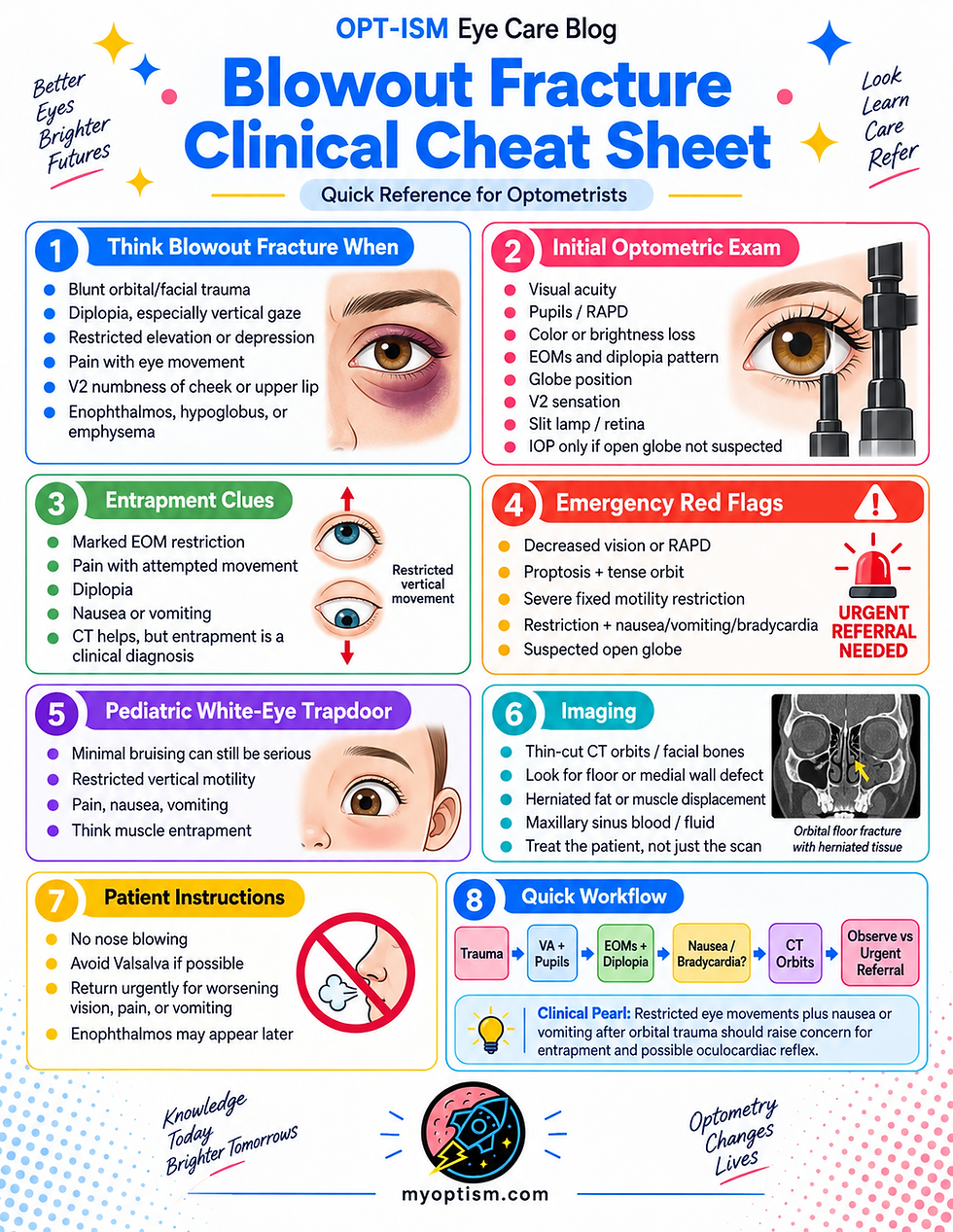
Blowout Fracture Clinical Cheat Sheet

Quick Reference for Optometrists
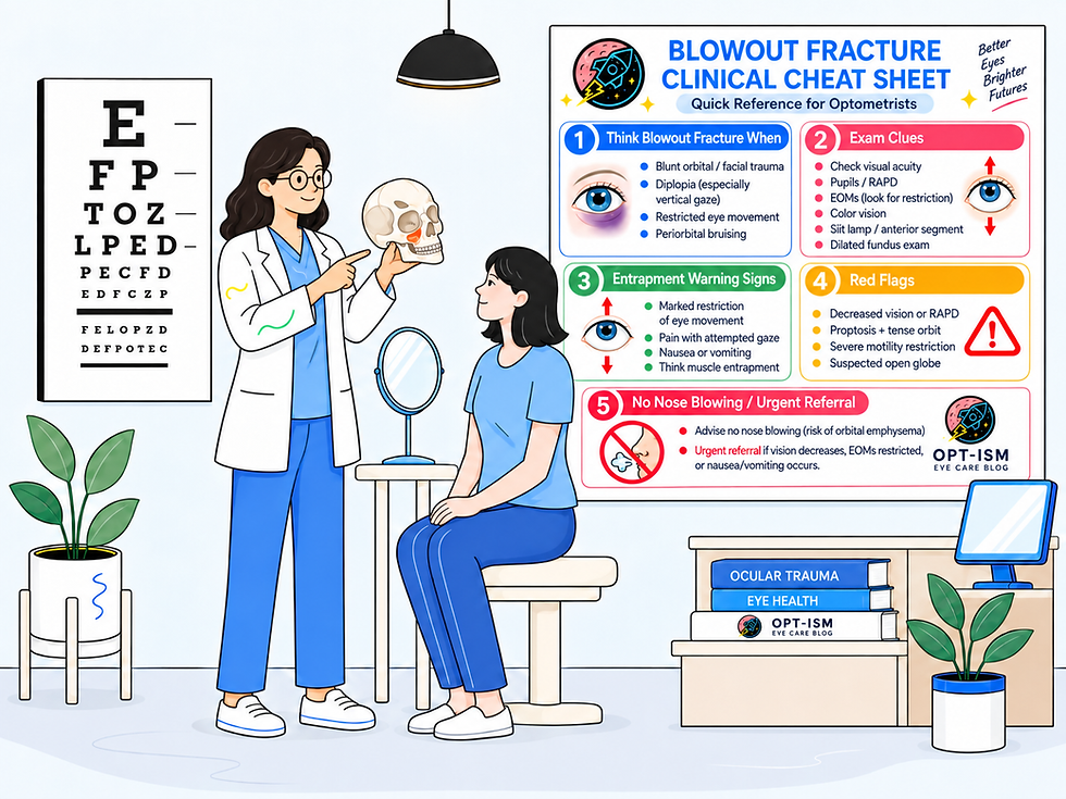
1. Think Blowout Fracture When You See
Blunt orbital/facial trauma + any of the following:
Diplopia, especially in vertical gaze
Restricted elevation or depression
Pain with eye movement
Periorbital edema/ecchymosis
Enophthalmos or hypoglobus
Infraorbital/V2 numbness of cheek, upper lip, or upper teeth
Orbital/subcutaneous emphysema
Nausea or vomiting associated with eye movement
The orbital floor and medial wall are the most commonly involved walls.
2. Optometric Trauma Exam
Check | Look For |
VA | Unexplained vision loss |
Pupils | RAPD |
Color/brightness | Traumatic optic neuropathy |
Confrontation VF | Optic nerve/neurologic defect |
External exam | Edema, ecchymosis, laceration, emphysema |
Globe position | Proptosis, enophthalmos, hypoglobus |
EOMs | Restriction—especially supraduction |
Diplopia | Primary gaze vs up/down gaze |
V2 sensation | Cheek/upper lip/upper teeth hypoesthesia |
Anterior segment | Abrasion, hyphema, iritis, open globe |
IOP | Only if open globe is not suspected |
Dilated retina | Commotio, tear, hemorrhage, RD |
Optic nerve | Traumatic optic neuropathy |
Clinical Pearl
Do not let the orbital fracture distract you from the globe or optic nerve.
3. Entrapment = Clinical Diagnosis
Suspect entrapment with:
Marked EOM restriction
pain with attempted movement
± diplopia
± nausea/vomiting
± bradycardia
CT may show herniated muscle or orbital fat, but radiographic herniation does not prove clinical entrapment—and significant clinical restriction can occur even when CT findings appear less dramatic.
Most Common
Orbital floor → inferior rectus / surrounding soft tissue
Medial Wall
Medial rectus involvement → horizontal restriction
4. The Oculocardiac Reflex 🚨
Think:
Restricted EOM + Nausea/Vomiting + Bradycardia
Entrapped orbital tissue or extraocular muscle can trigger a trigeminal-vagal reflex causing:
Bradycardia
Nausea
Vomiting
Dizziness/syncope
Hypotension
Heart block
Rarely severe cardiac instability
This warrants urgent surgical evaluation, particularly when persistent or associated with hemodynamic instability.
5. Pediatric White-Eye Blowout Fracture 🚨
Children and young adults may develop a trapdoor fracture in which flexible bone briefly opens and then snaps back, trapping orbital tissue.
Classic Pattern
Minimal bruising/swelling
marked vertical EOM restriction
pain
nausea/vomiting
Do not be reassured by a normal-looking external eye.
Think:
“White eye + restricted motility = possible trapdoor entrapment.”
Pediatric trapdoor fractures are particularly associated with oculocardiac symptoms.
6. Emergency Red Flags 🚨
Immediate ED / Ophthalmology / Orbital Surgery Evaluation
↓ VA or RAPD→ Open globe, traumatic optic neuropathy, retinal injury, orbital compartment syndrome
Proptosis + tense orbit + ↓ vision/RAPD ± high IOP→ Orbital compartment syndrome
Severe/fixed EOM restriction→ Entrapment
Restriction + nausea/vomiting/bradycardia→ Oculocardiac reflex
Child with restricted vertical motility→ Trapdoor fracture
Suspected open globe→ No tonometry or pressure on globe
7. Imaging
Imaging of Choice:
Thin-cut CT of the orbits/facial bones
AAO EyeWiki describes approximately 1.0–1.5 mm axial cuts with coronal reconstruction for suspected orbital floor fractures.
Look for:
Orbital floor/medial wall defect
Orbital fat herniation
Inferior or medial rectus displacement
Maxillary sinus blood/fluid
Orbital emphysema
Retrobulbar hemorrhage
Associated facial fractures
Remember:
Treat the patient, not just the CT.
8. Classic CT Finding
“Teardrop Sign”
Orbital soft tissue protrudes through the fractured orbital floor into the maxillary sinus.
It indicates herniation, but does not by itself establish clinical muscle entrapment.
9. Diplopia: Entrapment or Swelling?
More Suggestive of Edema/Contusion | More Suggestive of Entrapment |
Mild-moderate restriction | Severe/fixed restriction |
Improving over several days | Little/no improvement |
Mild discomfort | Significant pain with movement |
No vagal symptoms | Nausea/vomiting/bradycardia |
Adult comminuted fracture | Pediatric trapdoor pattern |
Serial motility measurements are extremely useful.
10. Document Motility Precisely
Instead of: “EOM restricted.”
Document: OD −3 supraduction with vertical diplopia beginning approximately 10° above primary gaze.
At follow-up: OD −1 supraduction — significant improvement from initial exam.
Prism measurements can also help objectively document improvement.
11. Patient Instructions
NO NOSE BLOWING
Communication between the orbit and sinus can allow air to enter the orbit.
AAO EyeWiki recommends avoiding nose blowing for approximately 4–6 weeks following an orbital floor fracture.
Also advise the patient to seek urgent care for worsening:
vision, diplopia, pain, proptosis, vomiting, restricted motility, or neurologic symptoms.
12. Observe vs Refer
Often Appropriate for Observation
Small/uncomplicated fracture with:
Stable vision
No RAPD
No entrapment
Mild diplopia
Improving motility
No troublesome enophthalmos
Surgical Evaluation
Consider for:
Persistent disabling diplopia
Motility that stops improving
Clinically significant enophthalmos/globe displacement
Significant orbital defect
Entrapment
Urgent/Emergent
Entrapment + severe restriction, severe pain, oculocardiac reflex, or hemodynamic instability.
13. Follow-Up
For adults with acute diplopia or pain with eye movement, AAO EyeWiki recommends reassessment at approximately 1 week, then serially while dysmotility continues to improve.
At each visit repeat:
VA → Pupils/RAPD → EOM → diplopia → globe position → V2 sensation → ocular health
Remember that enophthalmos may become more noticeable days to weeks later as edema resolves.
Blowout Fracture: 30-Second Clinical Workflow
TRAUMA
↓
VA + PUPILS/RAPD
↓
Vision loss / RAPD?→ 🚨 URGENT EVALUATION
↓
CHECK EOM + DIPLOPIA
↓
Marked restriction?→ Think ENTRAPMENT
↓
ASK ABOUT NAUSEA/VOMITING
CHECK HR IF CONCERNED
↓
Restriction + vagal symptoms/bradycardia?→ 🚨 OCULOCARDIAC REFLEX → URGENT SURGICAL EVALUATION
↓
CT ORBITS / FACIAL BONES
↓
NO NOSE BLOWING
↓
OBSERVE vs OCULOPLASTICS / ORBITAL SURGERY
↓
RECHECK MOTILITY + DIPLOPIA + ENOPHTHALMOS


Comments