
Orbital Blowout Fractures: What Optometrists Need to Know

A patient presents after being struck in the eye with a baseball, fist, elbow, airbag, or other blunt object. The eyelids are swollen, the patient reports double vision, and elevation of the eye appears restricted.
One diagnosis should immediately move toward the top of the differential:
Orbital blowout fracture.
Although orbital fractures are frequently managed by ophthalmology, oculoplastics, ENT, or maxillofacial surgery, the optometrist may be the first eye-care provider to evaluate the patient. Recognizing the difference between routine post-traumatic swelling and extraocular muscle entrapment, orbital compartment syndrome, traumatic optic neuropathy, or an open globe can be vision-saving—and occasionally life-saving.
This guide reviews the clinical features, examination, imaging, red flags, referral considerations, and follow-up principles optometrists should know.

What Is an Orbital Blowout Fracture?
A blowout fracture occurs when blunt trauma causes a fracture of one of the thin walls of the orbit while the orbital rim may remain relatively intact.
The orbital floor is the classic location, although the medial orbital wall can also fracture.
With an orbital floor fracture, orbital fat and other soft tissue may herniate through the defect into the maxillary sinus. In some cases, orbital tissue can become caught or tethered within the fracture.
The consequences may include:
Diplopia, restricted ocular motility, pain with eye movement, infraorbital nerve hypoesthesia, enophthalmos, orbital emphysema, and extraocular muscle or soft-tissue entrapment.
Large defects can increase orbital volume and eventually produce enophthalmos. Entrapped tissue can create a much more urgent problem.
Why Does the Orbit “Blow Out”?
Two major mechanisms have traditionally been proposed.
Hydraulic Mechanism
A sudden force directed at the globe dramatically increases intraorbital pressure. That pressure is transmitted to the thin orbital walls, causing the floor or medial wall to fracture.
Buckling Mechanism
Force applied to the orbital rim is transmitted posteriorly through the orbital bones, causing one of the thinner walls to buckle and fracture.
In real-world trauma, elements of both mechanisms may occur.
Common causes include sports injuries, physical assault, falls, motor vehicle accidents, bicycle accidents, and being struck by objects such as baseballs, racquetballs, or other projectiles.
The Classic Orbital Floor Fracture
The orbital floor separates the orbit from the maxillary sinus.
When it fractures, orbital fat can prolapse downward into the sinus. The inferior rectus and surrounding connective tissues may become displaced, tethered, or entrapped.
That anatomy explains one of the classic presentations:
Diplopia with limited elevation following blunt orbital trauma.
But an important clinical pearl is that restricted elevation does not automatically mean the inferior rectus is entrapped.
Restriction may also result from edema, hemorrhage, muscle contusion, pain, or mechanical tethering of adjacent orbital tissues.
Entrapment must therefore be interpreted from the entire clinical picture rather than CT appearance alone.
Symptoms Optometrists Should Ask About
History is extremely important.
Ask specifically about diplopia and whether it occurs in primary gaze, upgaze, downgaze, or another direction. Determine whether there is pain with eye movement, decreased vision, numbness of the cheek or upper lip, nausea or vomiting, dizziness or faintness, flashes or floaters, photophobia, epistaxis, or a subjective change in the position of the eye.
The mechanism matters as well.
Ask what struck the patient, where the impact occurred, when the injury happened, and whether there was loss of consciousness or other facial or head trauma.
Numbness involving the ipsilateral cheek, side of the nose, upper lip, or upper teeth may indicate involvement of the infraorbital nerve, a branch of V2, which travels along the orbital floor.
The Optometrist's Examination
The orbital fracture itself should never distract the clinician from evaluating the globe and optic nerve.
Orbital trauma may coexist with serious ocular injuries.
A systematic examination is essential.
Examination | What You Are Looking For |
Visual acuity | Unexplained reduction in vision |
Pupils | RAPD suggesting optic nerve or severe retinal injury |
Color vision/red desaturation | Possible traumatic optic neuropathy |
Confrontation fields | Gross optic nerve or neurologic deficit |
External examination | Ecchymosis, edema, laceration, asymmetry |
Globe position | Proptosis, enophthalmos, hypoglobus |
Extraocular movements | Restriction, particularly elevation |
Diplopia assessment | Direction and severity of binocular diplopia |
V2 sensation | Infraorbital nerve involvement |
Slit lamp | Abrasion, traumatic iritis, hyphema, globe injury |
IOP | Elevated pressure or orbital compartment syndrome when safe to measure |
Dilated fundus examination | Commotio, retinal tear, hemorrhage, detachment, optic nerve injury |
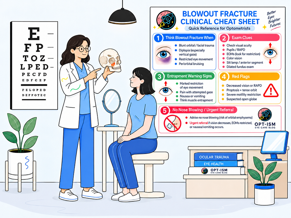
Important: If an open globe is suspected, avoid tonometry or anything that places pressure on the eye and arrange emergency ophthalmic evaluation.
Look at the Eye Movements Carefully
Extraocular motility is one of the most important parts of the examination.
With an orbital floor fracture, limitation of elevation is common.
Ask the patient whether attempted elevation causes:
Diplopia + pain + nausea
That combination should significantly increase suspicion for entrapment.
Do not simply record:
“EOM restricted.”
Document the actual pattern.
For example:
OD: approximately 50% limitation of supraduction with binocular vertical diplopia beginning 10 degrees above primary gaze.
That documentation becomes extremely useful when the patient is examined again several days later.
Prism measurements in primary gaze and affected fields of gaze can also help determine whether motility is improving over time. The AAO notes that progressively decreasing deviations can support continued observation in appropriate cases.
Entrapment Is a Clinical Diagnosis
This is one of the most important concepts for clinicians evaluating orbital fractures.
Do not depend entirely on the CT report to determine whether an extraocular muscle is entrapped.
CT may demonstrate a fracture, herniated orbital fat, displacement of the inferior rectus, or tissue extending into the maxillary sinus.
But radiologic herniation is not synonymous with clinical entrapment.
Conversely, significant clinical entrapment can sometimes exist without dramatic CT findings.
According to AAO EyeWiki, entrapment is fundamentally a clinical diagnosis.
The patient's motility, pain, diplopia, age, mechanism of injury, and systemic symptoms must all be considered.
The Pediatric “White-Eye Blowout Fracture”
Children deserve special attention.
Because pediatric bones are more flexible, the orbital floor can fracture and then partially spring back into position.
Imagine a hinged door briefly opening and then closing.
Soft tissue or an extraocular muscle can become caught when the bone returns toward its original position.
This is commonly called a:
Trapdoor fracture.
Unlike many adult orbital fractures, these injuries can have surprisingly little external bruising or swelling.
The child may therefore have a relatively normal-looking eye—the classic:
“White-eye blowout fracture.”
Do not let the lack of dramatic ecchymosis provide false reassurance.
A child with orbital trauma, restricted vertical eye movement, pain, nausea, or vomiting should raise immediate concern for entrapment.
The Oculocardiac Reflex: A Red Flag You Cannot Miss
One of the most clinically important complications of orbital tissue entrapment is the oculocardiac reflex.
Traction or pressure involving the extraocular muscles or orbital tissues can stimulate the trigeminal-vagal reflex pathway.
The patient may develop:
Bradycardia, nausea, vomiting, dizziness, syncope, hypotension, heart block, or—in severe circumstances—significant cardiac instability.
A particularly concerning presentation is:
Orbital trauma + restricted motility + nausea/vomiting + bradycardia.
This should not simply be attributed to pain, anxiety, or concussion.
Think:
Entrapment with oculocardiac reflex.
Urgent surgical evaluation is warranted because persistent entrapment may threaten the function of the involved muscle, and significant vagal responses can produce cardiovascular instability.
Do Not Forget Orbital Compartment Syndrome
Not every dangerous post-traumatic orbit involves a blowout fracture.
Retrobulbar hemorrhage can rapidly increase pressure within the orbit and produce orbital compartment syndrome.
This represents a separate ophthalmic emergency.
Be concerned when orbital trauma is followed by rapidly decreasing vision, an RAPD, marked proptosis, a tense orbit, severe pain, ophthalmoplegia, resistance to retropulsion, or markedly elevated IOP.
Visual loss may become permanent if optic nerve perfusion is compromised.
A patient with suspected orbital compartment syndrome requires immediate emergency ophthalmic management. The urgency should not be reduced simply because CT imaging has not yet been completed.
Traumatic Optic Neuropathy Must Also Be Considered
Orbital trauma can damage the optic nerve even when the globe initially appears relatively normal.
Look for a reduction in visual acuity that cannot be explained by the anterior segment, an RAPD, color desaturation, reduced brightness perception, or a visual field defect.
A normal-appearing optic disc does not exclude acute traumatic optic neuropathy because the injury may initially be retrobulbar.
An RAPD after orbital trauma should therefore never be dismissed.
CT Is the Imaging Study of Choice
When an orbital fracture is suspected, computed tomography is the primary imaging modality.
Thin-section CT of the orbits/facial bones with multiplanar reconstruction allows evaluation of the orbital walls, maxillary and ethmoid sinuses, extraocular muscles, orbital fat, hemorrhage, foreign bodies, and associated facial fractures. AAO references thin cuts of approximately 1.0–1.5 mm with coronal reconstruction for suspected orbital floor fractures.
CT may demonstrate orbital floor discontinuity, herniation of orbital fat into the maxillary sinus, displacement of the inferior rectus, maxillary sinus hemorrhage or fluid, orbital emphysema, or other orbital and facial fractures.
Again:
The scan supports the clinical examination. It does not replace it.
What About the “Teardrop Sign”?
Clinicians may encounter the term teardrop sign when discussing orbital floor fractures.
This refers to soft tissue hanging from the orbit into the maxillary sinus, creating a teardrop-like configuration on imaging.
It suggests herniation of orbital contents through the orbital floor.
However, the presence of herniated tissue on imaging alone does not prove that the inferior rectus muscle is clinically entrapped.
Correlate the scan with motility.
Enophthalmos May Appear Later
Immediately after trauma, significant edema and hemorrhage can mask changes in orbital volume.
The patient may even initially appear mildly proptotic.
As swelling resolves over subsequent days or weeks, enophthalmos may become more apparent.
Large orbital defects allow orbital tissue to displace into the adjacent sinus, effectively increasing the volume of the orbit.
This explains why assessment of globe position at follow-up is important even when the patient's initial appearance seems symmetrical.
When Does a Blowout Fracture Need Surgery?
Not every orbital floor fracture requires surgical repair.
Many uncomplicated fractures improve as edema resolves.
Indications are individualized, but surgical evaluation becomes increasingly important when there is severe or persistent restrictive diplopia, clinically significant enophthalmos or globe displacement, a large fracture with substantial orbital tissue displacement, or evidence of entrapment.
True entrapment with oculocardiac reflex or severe motility restriction is different from routine post-traumatic diplopia and warrants urgent evaluation.
Traditional teaching frequently mentions repair within two weeks. The reality is more nuanced.
AAO EyeWiki notes that many patients with less severe dysmotility improve during observation and that there is not compelling evidence for a universal mandatory “two-week window.” If motility continues to improve, surgical decision-making may be delayed. Persistent disabling diplopia without improvement after the initial observation period is more concerning.
For the optometrist, the important point is not deciding the exact surgical date.
The important point is recognizing which patient needs routine specialty follow-up and which patient needs urgent intervention.
Nose Blowing: A Small Instruction That Matters
Patients with orbital floor or medial wall fractures may have communication between the orbit and adjacent paranasal sinuses.
Tell these patients:
Do not blow your nose.
Forceful nose blowing can push air from the sinus into the orbit and produce or worsen orbital and subcutaneous emphysema.
AAO EyeWiki recommends avoiding nose blowing for approximately 4–6 weeks following an orbital floor fracture.
Patients should also avoid deliberately performing Valsalva-type maneuvers unless instructed otherwise by their treating specialist.
Steroids and Antibiotics
Some patients with significant orbital edema or dysmotility may receive a short course of systemic corticosteroids to determine how much motility restriction is related to swelling.
Antibiotics are sometimes prescribed, particularly when there is associated sinus disease or other risk factors.
However, these are not substitutes for proper evaluation of suspected entrapment, globe injury, optic neuropathy, or orbital compartment syndrome.
Medication decisions should be individualized and coordinated with the clinician managing the orbital fracture.
Practical Referral Categories for the Optometrist
Presentation | Suggested response |
Reduced vision, RAPD, suspected open globe | Emergency ophthalmology/ED evaluation |
Proptosis + vision loss + RAPD/very high IOP | Suspect orbital compartment syndrome — emergency |
Restricted EOM + nausea/vomiting/bradycardia | Suspect entrapment/OCR — urgent surgical evaluation |
Pediatric restricted motility with minimal bruising | Suspect white-eye trapdoor fracture — urgent |
Significant motility restriction or severe pain | Urgent ophthalmic/orbital evaluation |
Mild diplopia improving as edema resolves | Close specialty follow-up may be appropriate |
New/worsening enophthalmos | Oculoplastic/orbital surgical evaluation |
Stable fracture without ocular complications | Monitor according to specialist treatment plan |
Follow-Up Matters
For an uncomplicated adult orbital floor fracture with diplopia or pain on eye movement, AAO EyeWiki recommends reevaluation within approximately one week, followed by serial assessments while motility abnormalities persist and continue to improve.
At follow-up, repeat visual acuity, pupils, extraocular motility, diplopia measurements, globe position, anterior segment examination, and posterior segment evaluation as clinically indicated.
Do not simply document:
“Patient doing better.”
Document whether the actual restriction is improving.
For example:
Day 1: −3 supraduction OD
Day 7: −1 supraduction OD
That tells the consulting surgeon substantially more about the course of recovery.
A Practical Blowout Fracture Workflow
Rule out vision-threatening ocular injury first. Check visual acuity, pupils/RAPD, optic nerve function, anterior segment, and posterior segment. Avoid pressure on the eye if an open globe is suspected. Then assess globe position, V2 sensation, extraocular motility, diplopia, pain, and systemic symptoms such as nausea or vomiting. Obtain or arrange thin-section orbital CT when fracture is suspected. If there is severe motility restriction, pediatric trapdoor fracture, oculocardiac symptoms, visual loss, optic neuropathy, or orbital compartment syndrome, escalate urgently. Otherwise, coordinate ophthalmology/oculoplastics follow-up, instruct the patient not to blow the nose, and document motility carefully so improvement or deterioration can be identified at subsequent visits.
Clinical Pearl: Swelling vs Entrapment
One of the hardest decisions after orbital trauma is determining why an eye is not moving normally.
Think about it this way:
Swelling tends to improve. Entrapment mechanically prevents movement.
Edema, hemorrhage, muscle contusion, pain, cranial nerve injury, and loss of fusion can all cause diplopia following trauma.
That is why serial examinations can be so valuable.
A patient whose ocular motility noticeably improves over several days behaves differently from a patient with severe, fixed restriction accompanied by pain, nausea, or bradycardia.
Five Findings You Should Never Miss
After orbital trauma, immediately increase your level of concern when you see:
Decreased vision or RAPD — consider optic neuropathy, globe injury, retinal injury, or orbital compartment syndrome.
Marked proptosis with a tense orbit — consider retrobulbar hemorrhage and orbital compartment syndrome.
Restricted ocular motility with nausea or vomiting — consider muscle or soft-tissue entrapment and the oculocardiac reflex.
A child with severe motility restriction but little bruising — consider a white-eye trapdoor fracture.
Progressive enophthalmos or persistent functionally significant diplopia — arrange orbital surgical evaluation.
The Bottom Line for Optometrists
Orbital blowout fractures are not simply “broken bones around the eye.”
The fracture itself may be relatively uncomplicated while the structures inside the orbit are not.
For optometrists, the priorities are to:
Protect the globe. Evaluate the optic nerve. Examine motility carefully. Recognize entrapment. Recognize the oculocardiac reflex. Recognize orbital compartment syndrome. Obtain appropriate imaging. Refer urgently when red flags are present.
And remember perhaps the most important clinical pearl:
A normal-looking CT does not completely rule out clinically significant entrapment—and a normal-looking external eye does not rule out a serious pediatric trapdoor fracture.
When trauma produces diplopia, restricted motility, pain, nausea, vomiting, bradycardia, reduced vision, or an RAPD, the examination needs to move beyond simply asking whether the orbital floor is fractured.
The real question is:
What happened to the eye, optic nerve, and orbital tissues because of the fracture?
That is where the optometrist can make the greatest difference.
References
American Academy of Ophthalmology EyeWiki. Orbital Floor Fractures.
American Academy of Ophthalmology EyeWiki. Oculocardiac Reflex.
American Academy of Ophthalmology EyeWiki. Orbital Medial Wall Fractures.
NCBI Bookshelf/StatPearls. Imaging of Ocular Trauma.
NCBI Bookshelf/StatPearls. Orbital Floor Fracture.
NCBI Bookshelf/StatPearls. Extraocular Muscle Management With Orbital and Globe Trauma.

Comments