
Eye Coloboma: 5-Question Multiple-Choice Quiz

Updated: Sep 16

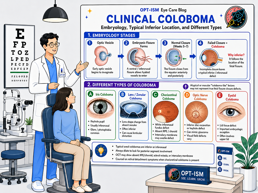
1. Why are typical ocular colobomas most commonly located inferiorly or inferonasally?
A. The inferior retina develops later than the superior retina
B. The embryonic fissure is located along the ventral/inferonasal aspect of the developing optic cup
C. Inferior ocular tissues receive less blood flow during development
D. The optic nerve migrates inferiorly during gestation
2. Which finding is most characteristic of a classic iris coloboma?
A. Superior sectoral iris atrophy
B. Diffuse absence of iris tissue
C. Inferonasal keyhole-shaped pupil
D. Central irregular pupil with posterior synechiae
3. Which OCT finding may be seen in a chorioretinal coloboma?
A. Uniform thickening of the RPE and choroid
B. Absence or severe thinning of the RPE and choroid with an intercalary membrane
C. Isolated vitreous hemorrhage without retinal abnormalities
D. Diffuse macular edema in every affected eye
4. What is one of the most important long-term complications of chorioretinal coloboma?
A. Acute angle-closure glaucoma
B. Retinal detachment
C. Central retinal artery occlusion
D. Corneal ectasia
5. Which is the BEST clinical approach when evaluating a patient with ocular coloboma?
A. Monitor the iris appearance only if visual acuity is good
B. Diagnose glaucoma whenever the optic nerve appears deeply excavated
C. Obtain a dilated examination and consider baseline widefield imaging, OCT, and visual field testing
D. Refer every patient immediately for prophylactic retinal laser
Answer Key
1. B | 2. C | 3. B | 4. B | 5. C
Correct answer: B
Typical colobomas follow the location of the embryonic fissure, which normally closes during approximately weeks 5–7 of gestation.
Correct answer: C
A typical iris coloboma produces an inferonasal keyhole-shaped pupil. Finding one should prompt a dilated examination to evaluate for posterior segment involvement.
Correct answer: B
OCT may demonstrate absent or markedly thinned RPE and choroid, scleral excavation or ectasia, and an overlying intercalary membrane. Imaging through the coloboma margin may reveal breaks, schisis-like changes, or fluid.
Correct answer: B
Retinal detachment is a major concern. Breaks may occur within the coloboma, along its margin, or elsewhere in the peripheral retina, so patients should be educated about flashes, floaters, a curtain or shadow, and sudden vision changes.
Correct answer: C
Coloboma evaluation should extend beyond the visible iris defect. Baseline imaging can document posterior segment anatomy, while congenital optic nerve excavation should not be mistaken for glaucoma without evidence of progression or other supporting findings.
Comments