
Overview From the Retina to the Visual Cortex

A Clinician-Friendly Guide to Image Formation, Retinal Signal Processing, and the Visual Pathway

Vision feels instant, but it is actually a highly organized neurologic process. Light enters the eye, is focused onto the retina, converted into electrical signals, refined by retinal circuitry, and then transmitted through the optic nerve and visual pathway to the visual cortex.
These three clinical diagrams work together to explain the full pathway of sight:
Image formation in the eye
Neurosensory retina and retinal signal processing
Visual pathway to the cortex and lesion localization
Understanding this pathway helps clinicians localize disease, interpret visual field defects, and explain vision to patients in a clear, meaningful way.

1. Image Formation: How Light Becomes a Retinal Image
Vision begins when light reflects off an object and enters the eye through the tear film, cornea, aqueous humor, pupil, lens, and vitreous before reaching the retina.
The cornea provides most of the eye’s focusing power, while the crystalline lens fine-tunes focus through accommodation. Together, these structures focus light onto the retina, creating a real, inverted image.
Key optical structures include:
Tear film: Creates a smooth refractive surface.
Cornea: Provides the majority of refractive power.
Anterior chamber: Contains aqueous humor and supports intraocular pressure.
Pupil and iris: Regulate the amount of light entering the eye.
Crystalline lens: Adjusts focus for near and far vision.
Vitreous: Maintains ocular shape and transmits light to the retina.
Retina: Converts the focused image into neural information.
A clinically important point is that the image projected onto the retina is spatially reversed. The superior visual field projects to the inferior retina, and the inferior visual field projects to the superior retina. The temporal visual field projects to the nasal retina, and the nasal visual field projects to the temporal retina.
This retinotopic organization is preserved throughout the visual pathway.

2. The Neurosensory Retina: Where Light Becomes a Neural Signal
The retina is neural tissue. It is not simply a screen at the back of the eye; it is an extension of the central nervous system that detects, converts, and processes visual information.
The retina can be thought of in two major functional zones:
Outer Retina: Sensory Detection
The outer retina contains the photoreceptors and their supporting structures. This is where light detection begins.
Important outer retinal structures include:
Retinal pigment epithelium, or RPE: Absorbs scattered light, supports photoreceptor metabolism, recycles visual pigment, and helps maintain the blood-retina barrier.
Photoreceptor outer segments: Contain photopigment discs where phototransduction begins.
Photoreceptor inner segments: Contain mitochondria and metabolic machinery.
Outer nuclear layer: Contains rod and cone nuclei.
Outer plexiform layer: Synaptic layer between photoreceptors, bipolar cells, and horizontal cells.
The choroid lies external to the retina and provides vascular support to the outer retina, especially the photoreceptors and RPE.
Inner Retina: Signal Processing
The inner retina receives signals from photoreceptors and begins refining the visual message before it leaves the eye.
Important inner retinal structures include:
Inner nuclear layer: Contains bipolar, horizontal, amacrine, and Müller cell bodies.
Inner plexiform layer: Synaptic layer where bipolar, amacrine, and ganglion cells interact.
Ganglion cell layer: Contains retinal ganglion cell bodies.
Retinal nerve fiber layer: Contains ganglion cell axons that converge to form the optic nerve.
Internal limiting membrane: The innermost retinal boundary formed by Müller cell endfeet.
This organization is essential for understanding OCT interpretation, retinal disease localization, and glaucoma-related nerve fiber loss.

3. Rods and Cones: Peripheral Vision, Color, and Acuity
The retina contains two major photoreceptor types: rods and cones.
Rods
Rods are highly sensitive to light and are most important for:
Dim-light vision
Peripheral vision
Motion detection
Night vision
Rods are concentrated more heavily in the peripheral retina and do not provide color vision.
Cones
Cones are responsible for:
Color vision
Fine detail
High-acuity central vision
Daylight vision
Cones are densely packed in the macula, especially at the fovea, where visual acuity is highest. The fovea is specialized for precise central vision because it has a high cone density and fewer overlying inner retinal layers.
This explains why macular disease affects reading, facial recognition, and fine detail, while peripheral retinal disease may affect side vision or night vision.

4. Phototransduction: How Photoreceptors Convert Light into Signal
Phototransduction is the biochemical process that allows rods and cones to convert light into neural signals.
In simplified form:
A photon strikes photopigment in the photoreceptor outer segment.
Opsin is activated.
Transducin is activated.
Phosphodiesterase reduces cGMP levels.
cGMP-gated sodium and calcium channels close.
The photoreceptor hyperpolarizes.
Glutamate release decreases.
This is a key concept because photoreceptors behave differently than many other neurons.
In the dark, photoreceptors are relatively depolarized and continuously release glutamate.In the light, photoreceptors hyperpolarize and release less glutamate.
That change in glutamate release is the beginning of visual signaling.

5. Inner Retinal Processing: ON, OFF, Horizontal, Amacrine, and Ganglion Cells
The retina does not simply passively transmit information. It actively processes contrast, edges, motion, and spatial relationships before the signal reaches the brain.
Bipolar Cells
Bipolar cells transmit signals from photoreceptors to ganglion cells.
There are two major functional types:
ON bipolar cells: Respond to light increments.
OFF bipolar cells: Respond to light decrements.
This ON/OFF organization helps the visual system detect contrast and changes in illumination.
Horizontal Cells
Horizontal cells provide lateral inhibition between photoreceptors. This contributes to center-surround receptive fields, which improve contrast sensitivity and edge detection.
Amacrine Cells
Amacrine cells modify signal timing, motion sensitivity, directionality, and complex retinal processing. They play an important role in dynamic visual function.
Retinal Ganglion Cells
Ganglion cells are the output neurons of the retina. Unlike photoreceptors and bipolar cells, which primarily use graded potentials, retinal ganglion cells generate action potentials.
Their axons form the retinal nerve fiber layer, converge at the optic disc, and become the optic nerve.
This is why glaucoma, optic neuropathy, and other retinal ganglion cell disorders can produce characteristic visual field defects.

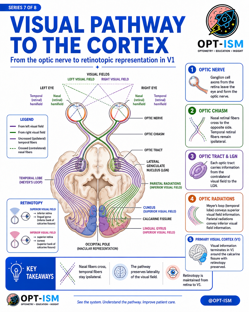
6. From Retina to Brain: The Visual Pathway
Once visual signals leave the eye, they travel through a highly organized pathway:
Retina
Optic nerve
Optic chiasm
Optic tract
Lateral geniculate nucleus, or LGN
Optic radiations
Primary visual cortex, or V1
At the optic chiasm, nasal retinal fibers cross, while temporal retinal fibers remain uncrossed. This crossing pattern allows the right brain to process the left visual field and the left brain to process the right visual field.
After the chiasm, visual information is organized by visual field, not by eye.
This is why post-chiasmal lesions often create homonymous visual field defects.
7. Optic Radiations and the Visual Cortex
After synapsing in the LGN, visual information travels through the optic radiations to the occipital cortex.
There are two major optic radiation pathways:
Temporal Lobe: Meyer’s Loop
Meyer’s loop carries information from the superior visual field. Lesions here can produce a contralateral superior quadrantanopia, often described as “pie in the sky.”
Parietal Radiations
Parietal radiations carry information from the inferior visual field. Lesions here can produce a contralateral inferior quadrantanopia, often described as “pie on the floor.”
Primary Visual Cortex
The primary visual cortex is located around the calcarine fissure in the occipital lobe.
Important cortical relationships include:
Cuneus: Represents the inferior visual field.
Lingual gyrus: Represents the superior visual field.
Occipital pole: Represents macular vision.
Macular sparing in a homonymous hemianopia may suggest occipital cortex involvement because the macular region often has dual or robust vascular representation.

8. Clinical Localization: Matching Lesions to Visual Field Defects
Visual field defects help clinicians localize pathology along the visual pathway.
Prechiasmal Lesions
A lesion before the chiasm, such as optic neuritis or compressive optic neuropathy, often causes monocular visual loss.
Chiasmal Lesions
A midline chiasmal lesion affects crossing nasal retinal fibers and classically causes bitemporal hemianopia.
This pattern raises concern for sellar or parasellar pathology, such as pituitary enlargement.
Optic Tract or Retrochiasmal Lesions
Lesions behind the chiasm cause contralateral homonymous visual field defects.
The more posterior the lesion, the more congruous the defect tends to be.
Temporal Lobe Lesions
Temporal lobe involvement affecting Meyer’s loop can cause a contralateral superior quadrantanopia.
Parietal Lobe Lesions
Parietal radiation involvement can cause a contralateral inferior quadrantanopia.
Occipital Cortex Lesions
Occipital cortex lesions can cause a contralateral homonymous hemianopia, often highly congruous and sometimes with macular sparing.
High-Yield Clinical Pearls
The eye forms a focused, inverted image on the retina, but the brain interprets that signal into upright visual perception.
The retina is neural tissue and begins processing visual information before the signal ever reaches the brain.
Rods dominate dim-light and peripheral vision, while cones dominate color, detail, and central acuity.
Phototransduction begins in the photoreceptor outer segments and results in decreased glutamate release in response to light.
Horizontal cells enhance contrast through lateral inhibition.
Amacrine cells refine timing, motion, and directional sensitivity.
Ganglion cells generate action potentials and form the optic nerve.
Nasal retinal fibers cross at the optic chiasm, while temporal retinal fibers remain ipsilateral.
Post-chiasmal lesions produce contralateral homonymous visual field defects.
Temporal lobe lesions affect the superior visual field, while parietal lesions affect the inferior visual field.
Why This Matters Clinically
Understanding the visual system helps clinicians connect anatomy with symptoms.
A patient with blurred central vision may have macular disease.A patient with peripheral field loss may have glaucoma, retinal disease, or neurologic involvement.A patient with a bitemporal field defect may need evaluation for chiasmal compression.A patient with a homonymous defect may require neuroimaging or neurologic workup.
The retina, optic nerve, and brain are deeply connected. Careful examination of the eyes can reveal not only ocular disease, but also neurologic and systemic disease.
Conclusion
Vision is a coordinated process involving optics, retinal neurobiology, and cortical interpretation. Light must be properly focused, detected by photoreceptors, processed by retinal circuitry, transmitted through the optic nerve and chiasm, relayed through the LGN, and finally interpreted by the visual cortex.
For clinicians, this pathway is essential for understanding visual field defects, retinal disease, optic neuropathy, glaucoma, and neuro-ophthalmic localization.
At OPT-ISM Eye Care in South Tampa, advanced testing such as retinal imaging, OCT, and visual field testing helps evaluate the retina, optic nerve, and visual pathway so patients receive a clearer understanding of their eye health and vision.


Comments