
Bergmeister Papilla: A Persistent Hyaloid Remnant at the Optic Nerve

A Bergmeister papilla is an uncommon congenital finding that may be discovered incidentally during a routine dilated eye examination or while reviewing optic nerve OCT imaging.
It represents a remnant of the fetal hyaloid vascular system, a temporary blood supply that plays an important role in development of the eye before birth. In most people, this vascular system regresses before birth. When a small portion persists near the optic nerve, it may appear as a Bergmeister papilla.
Although the finding is usually benign and does not require treatment, recognizing its characteristic appearance is important because it can sometimes resemble optic disc pathology, including neovascularization, gliosis, or other anomalous tissue.

What Is a Bergmeister Papilla?
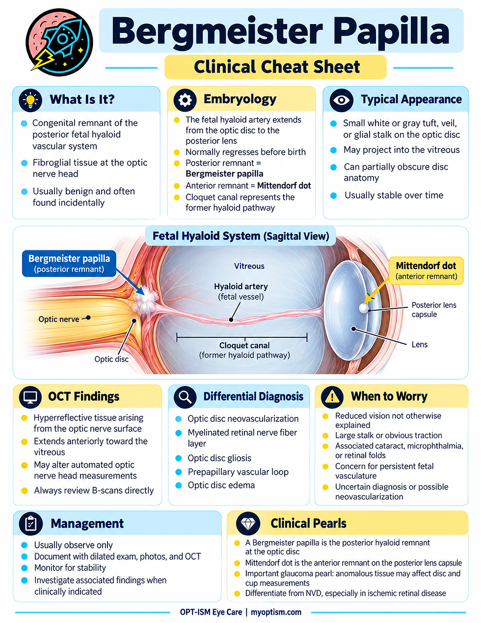
A Bergmeister papilla is a congenital remnant of the posterior fetal hyaloid system located at the optic nerve head.
Clinically, it may appear as a small tuft, membrane, stalk, or veil of whitish-gray fibroglial tissue extending anteriorly from the optic disc into the vitreous.
It is sometimes referred to as an epipapillary veil.
The tissue represents persistence of the fibrous or glial sheath that once surrounded the fetal hyaloid artery.
In many patients, the finding is subtle and may not be recognized until high-resolution OCT imaging is performed.
Understanding the Fetal Hyaloid System
To understand a Bergmeister papilla, it helps to understand how the fetal eye receives blood during development.
Early in development, the crystalline lens and portions of the developing eye require their own temporary vascular supply.
The hyaloid artery travels from the optic nerve through the vitreous toward the posterior surface of the developing lens. It then contributes to a network of blood vessels surrounding the lens known as the tunica vasculosa lentis.
As the eye matures and the retinal circulation develops, the hyaloid vascular system is no longer needed.
The system normally undergoes progressive involution during fetal development, leaving behind the largely transparent pathway known as Cloquet's canal within the vitreous.
If portions of the fetal system fail to completely regress, several recognizable ocular findings may remain.
Bergmeister Papilla vs. Mittendorf Dot
Two classic remnants of the fetal hyaloid system are particularly important for eye-care providers to recognize.
Bergmeister Papilla
The posterior remnant occurs at the optic nerve head and is called a Bergmeister papilla.
Mittendorf Dot
The anterior remnant occurs on the posterior lens capsule and is called a Mittendorf dot.
A Mittendorf dot typically appears as a small opacity located slightly inferonasal to the center of the posterior lens capsule.
Conceptually:
Optic nerve → hyaloid artery → vitreous → posterior lens
The remnants can therefore be remembered as:
Posterior remnant at the optic disc = Bergmeister papilla
Anterior remnant at the lens = Mittendorf dot
Both can occur as manifestations of incomplete regression of the fetal hyaloid system.
What Does a Bergmeister Papilla Look Like?
The appearance can vary considerably.
A Bergmeister papilla may appear as:
A small white or gray tuft on the optic nerve
A thin translucent membrane
A veil-like structure extending over part of the disc
A fibroglial stalk projecting anteriorly into the vitreous
A small area of tissue covering the central optic nerve
Tissue extending from the central or nasal portion of the disc
The finding may be unilateral or bilateral.
In some eyes, the remnant may partially obscure the underlying optic nerve anatomy.
What Does It Look Like on OCT?
Optical coherence tomography can be extremely helpful when evaluating a suspected Bergmeister papilla.
On OCT, the lesion may appear as hyperreflective tissue arising from the surface of the optic nerve and extending anteriorly toward the vitreous cavity.
Depending on its size and orientation, the tissue may resemble a small stalk, membrane, or triangular structure attached to the optic nerve.
OCT is particularly useful because it can help demonstrate that the structure originates from the optic nerve surface rather than representing edema within the nerve itself.
Modern OCT imaging may actually identify subtle Bergmeister papillae that are difficult to appreciate ophthalmoscopically.
Clinical Pearl: Bergmeister Papilla Can Affect OCT Measurements
One particularly important consideration involves optic nerve OCT interpretation.
Because the tissue projects from the optic nerve head, it can influence automated measurements of the optic disc and optic cup.
A prospective study evaluating eyes with Bergmeister papilla found that the anomaly can influence certain optic nerve head measurements obtained with spectral-domain OCT.
This becomes especially relevant when evaluating a patient for glaucoma.
For example, automated segmentation may interpret the tissue as part of the disc anatomy, potentially altering parameters such as:
Cup-to-disc measurements
Rim measurements
Disc morphology
Optic nerve head segmentation
Therefore, clinicians should always inspect the actual OCT B-scans rather than relying exclusively on automated numerical values.
Is a Bergmeister Papilla Dangerous?
In most patients, no.
An isolated Bergmeister papilla is usually a benign congenital finding and is frequently discovered incidentally.
Most patients have no symptoms and require no treatment.
However, its clinical significance depends on whether it is truly an isolated remnant or part of a more extensive developmental abnormality.
Relationship to Persistent Fetal Vasculature
More extensive persistence of the fetal hyaloid vascular system is known as persistent fetal vasculature, or PFV.
PFV was historically called persistent hyperplastic primary vitreous, or PHPV.
Unlike a small isolated Bergmeister papilla, significant PFV may involve:
Persistent hyaloid vessels
Fibrovascular stalks
Retrolental membranes
Cataract
Elongated ciliary processes
Vitreoretinal traction
Retinal folds
Retinal detachment
Microphthalmia
Posterior manifestations of persistent fetal vasculature may include a Bergmeister papilla, a persistent vascular stalk, retinal folds, or retinal detachment.
Therefore, when the finding appears unusually prominent, particularly in a child or an eye with reduced vision, the remainder of the eye should be carefully evaluated for evidence of more extensive PFV.
Differential Diagnosis
Recognizing a Bergmeister papilla is important because several conditions can create abnormal tissue at or near the optic nerve.
The differential diagnosis may include:
Optic Disc Neovascularization
Neovascularization of the disc, or NVD, may sometimes resemble fibrovascular tissue extending from the optic nerve.
This distinction is particularly important in patients with conditions such as:
Proliferative diabetic retinopathy
Retinal vein occlusion
Ocular ischemic syndrome
Other ischemic retinal disease
Unlike a stable congenital Bergmeister papilla, neovascularization is typically associated with retinal vascular pathology.
Fluorescein angiography can be useful when the diagnosis is uncertain because active neovascular tissue generally demonstrates leakage.
Prepapillary Vascular Loop
Occasionally, vessels may extend anteriorly from the optic disc and loop back toward the retina.
Some Bergmeister papillae can also contain or be associated with persistent vascular tissue, making careful examination important.
Optic Disc Gliosis
Gliotic tissue may create a whitish appearance on the optic nerve and can occasionally resemble a congenital remnant.
Myelinated Retinal Nerve Fiber Layer
Myelinated nerve fibers are typically opaque white lesions extending along the retinal nerve fiber layer from or near the optic disc.
Their feathered margins and distribution generally distinguish them from a Bergmeister papilla.
Optic Disc Edema
A Bergmeister papilla should not be mistaken for papilledema or other causes of optic nerve swelling.
Careful examination of:
Disc margins
Peripapillary RNFL
Retinal vessels
OCT B-scans
Symptoms
Fellow eye
usually helps distinguish these conditions.
When Is Additional Imaging Helpful?
Most isolated cases can be diagnosed clinically.
Additional testing may be useful when the diagnosis is uncertain.
OCT
OCT can demonstrate the structure of the tissue and its relationship to the optic nerve.
Fundus Photography
Baseline photography is useful for documenting the appearance and confirming long-term stability.
OCT Angiography
OCT-A may help determine whether vascular flow is present within a suspected persistent vascular remnant.
Fluorescein Angiography
Fluorescein angiography may be considered when optic disc neovascularization is part of the differential diagnosis.
A nonvascular Bergmeister papilla should not demonstrate the characteristic leakage expected from active neovascularization.
Does It Affect Vision?
An isolated Bergmeister papilla typically does not significantly affect visual acuity.
Vision problems should therefore prompt evaluation for another associated condition rather than automatically attributing the decreased vision to the papilla itself.
Possible associated abnormalities may include:
Persistent fetal vasculature
Retinal traction
Macular involvement
Cataract
Amblyopia
Other developmental abnormalities
The clinical context matters greatly.
Management
For an isolated, asymptomatic Bergmeister papilla, treatment is generally unnecessary.
Management typically consists of:
Documenting the finding.
Obtaining baseline photography or OCT when appropriate.
Evaluating the surrounding optic nerve and retina.
Confirming that there are no associated abnormalities.
Monitoring during routine comprehensive eye examinations.
A stable congenital remnant generally requires observation rather than intervention.
Clinical Pearls for Optometrists
Look at the B-scan.
Do not rely entirely on automated optic nerve OCT measurements when unusual tissue is present on the disc.
Remember the embryology.
Bergmeister papilla represents the posterior remnant of the fetal hyaloid system.
Look at the lens.
A Mittendorf dot may provide another clue to persistence of the fetal hyaloid system.
Differentiate it from NVD.
This is particularly important in diabetic patients or anyone with significant retinal ischemia.
Consider PFV in unusual cases.
A large stalk, abnormal lens, microphthalmia, retinal traction, or decreased vision should prompt consideration of more extensive persistent fetal vasculature.
Document stability.
Fundus photography and OCT provide useful baseline documentation.
Bergmeister Papilla vs. Important Look-Alikes
Finding | Typical Location | Key Feature |
Bergmeister papilla | Optic nerve head | Congenital fibroglial hyaloid remnant |
Mittendorf dot | Posterior lens capsule | Anterior remnant of fetal hyaloid system |
Persistent hyaloid artery | Optic disc toward posterior lens | Persistent vascular or fibrous stalk |
NVD | Optic disc | Abnormal new vessels, often associated with retinal ischemia |
Myelinated nerve fibers | Peripapillary retina | White, feathered RNFL appearance |
Optic disc edema | Optic nerve head | Disc elevation with swelling and associated clinical signs |
The Bottom Line
A Bergmeister papilla is a congenital remnant of the fetal hyaloid vascular system located at the optic nerve head.
In most patients, it is harmless and discovered incidentally during an eye examination.
The most important clinical considerations are recognizing its characteristic appearance, understanding its embryologic origin, distinguishing it from optic disc neovascularization and other pathology, and recognizing when a more extensive form of persistent fetal vasculature may be present.
OCT and fundus photography can be particularly useful for confirming the anatomy and documenting stability.
For eye-care providers, recognizing this unusual but generally benign optic nerve finding can prevent unnecessary concern while ensuring that important retinal and developmental conditions are not overlooked.


Comments