
How We See: A Simple Guide to How the Eye Forms an Image

Most people think we see with our eyes, but that is only partly true. Your eyes collect and focus light, but your brain is what turns those light signals into the image you understand.
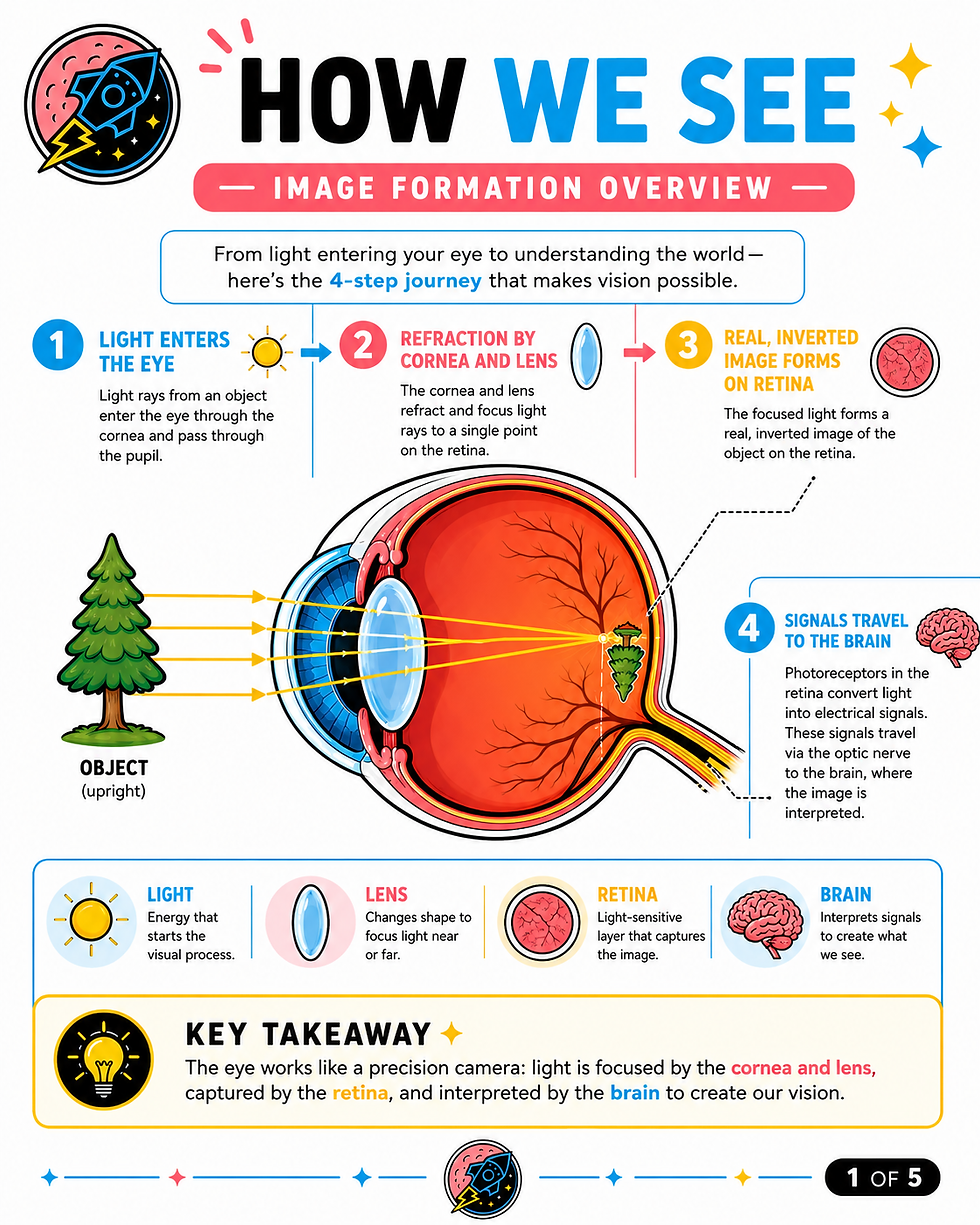
This image shows the basic steps of how vision works: light enters the eye, bends through the front structures, focuses on the retina, travels through the optic nerve, and is interpreted by the brain. The National Eye Institute describes this same process: light passes through the cornea and pupil, focuses through the lens onto the retina, and then electrical signals travel through the optic nerve to the brain.
Step 1: Light Enters the Eye
Vision starts when light reflects off an object. In the image, that object is a tree.
The light rays travel toward the eye and first pass through the cornea, which is the clear front surface of the eye. The cornea acts like the eye’s front window and helps bend light so it can be focused.
After passing through the cornea, light enters through the pupil. The pupil is the black opening in the center of the eye. The colored part of the eye, called the iris, controls the size of the pupil and decides how much light gets in. In bright light, the pupil gets smaller. In dim light, it gets larger.

Step 2: The Cornea and Lens Focus the Light
After light enters the eye, it passes through the lens.
The cornea and lens work together like a camera lens. Their job is to bend, or refract, light so it lands clearly on the back part of the eye called the retina.
The lens can also change shape slightly to help you focus at different distances. This is why younger eyes can usually shift focus from far away to up close more easily. As we age, the lens becomes less flexible, which is one reason many people need reading glasses after age 40.

Step 3: The Image Forms on the Retina
The retina is the light-sensitive tissue lining the back of the eye. In the image, the tree is focused onto the retina.
One important detail: the image that lands on the retina is actually upside down and reversed. That sounds strange, but your brain automatically corrects the image so you see the world upright.
The retina contains special light-sensing cells called photoreceptors. There are two main types:
Rods help you see in dim light and are important for night vision.
Cones help you see color and fine detail, especially in brighter lighting.
When light reaches the retina, these cells convert light into electrical signals that can be sent to the brain.

Step 4: The Optic Nerve Sends the Signal to the Brain
Once the retina converts light into electrical signals, those signals travel through the optic nerve.
The optic nerve is like a cable connecting the eye to the brain. It carries visual information from the retina toward the visual centers of the brain. The National Eye Institute describes the optic nerve as a bundle of more than 1 million nerve fibers that carry visual messages from the retina to the brain.

Step 5: The Brain Creates the Image You See
The final step happens in the brain.
Your brain receives the electrical signals from both eyes and organizes them into the image you experience. This is why vision is not just an eye function — it is an eye-and-brain function.
Your eyes capture the light, but your brain gives the image meaning.

Why This Matters During an Eye Exam
A comprehensive eye exam checks more than just whether you need glasses.
Your eye doctor evaluates several parts of the visual system, including:
The cornea, which helps focus light.
The pupil, which controls how much light enters.
The lens, which helps focus images clearly.
The retina, which captures light and begins the signal process.
The optic nerve, which carries visual information to the brain.
This is why eye exams can detect more than blurry vision. They can also reveal signs of cataracts, retinal disease, glaucoma, optic nerve problems, and other medical conditions that may affect how visual information travels from the eye to the brain.
Key Takeaway
The eye works like a precision camera, but vision is more advanced than a camera. Light enters the eye, gets focused onto the retina, turns into electrical signals, travels through the optic nerve, and is interpreted by the brain.
In simple terms:
The eye captures the image. The retina converts it. The optic nerve carries it. The brain makes sense of it.


Comments